Machine learning, imaging technique provide better insight into colorectal tissue
A team from McKelvey Engineering and the School of Medicine has developed a new method to detect cancerous tissue in the colon by combining machine learning and imaging

A team of biomedical engineers and colorectal surgeons at Washington University in St. Louis has used a new machine-learning algorithm coupled with a new hand-held multi-wavelength imaging device to better differentiate abnormal from normal colorectal tissues. The method may improve colorectal cancer screening, surveillance and treatment decision-making in the future.
Definitive screening for colon cancer and post-treatment rectal cancer surveillance currently relies on visual inspection of tissue with an endoscopic camera. This technique cannot detect subsurface lesions and lacks quantitative measures, limiting its accuracy. To better detect cancer in early stages of growth or recurrence, clinicians need improved imaging modalities.
Shuying Li and Yifeng Zeng, both doctoral students in the lab of Quing Zhu, professor of biomedical engineering in the McKelvey School of Engineering, and William C. Chapman Jr., MD, a general surgery resident working with Matthew Mutch, MD, chief of colorectal surgery at the School of Medicine, used the new hand-held multiwavelength spatial frequency domain imaging (SFDI probe to image resected colon tissue from 16 human patients. A novel AdaBoost machine-learning algorithm using tissue absorption and scattering features was then developed for automatic tissue classification. Using a total of 88 regions of interest (44 normal areas, 14 benign polyps and 30 cancer areas) collected in this study, the authors demonstrated excellent model classification. A superior area under the receiver operating characteristic curve of 0.95 was achieved for distinguishing between normal and abnormal colorectal tissue, Zhu said.
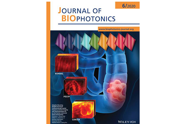
The results were published in the June issue of the Journal of Biophotonics with an image featured on the inside cover. The team is the first to use an AdaBoost-based multi-wavelength SFDI system to classify colorectal tissues, including normal, cancerous, and benign polyps. Potential applications of this technology include incorporation with traditional flexible endoscopy devices or creation of novel rigid proctoscopy platform.
The study was funded by the National Institutes of Health (R01 CA228047, R01 EB002136 and T32CA009621).
Li S, Zeng Y, Chapman W, Erfanzadeh M, Nandy S, Mutch M, Zhu Q. Adaptive Boosting (AdaBoost)‐based multiwavelength spatial frequency domain imaging and characterization for ex vivo human colorectal tissue assessment. Journal of Biophotonics, June 2020. Published online March 3, 2020. https://doi.org/10.1002/jbio.201960241


