Team developing tool to handle big data generated by advanced imaging tool
Washington University collaboration aims to advance 3D imaging of living cells, tissues
Light-sheet fluorescence microscopy, an imaging tool that can rapidly produce 3D images of complex cellular structures, gives scientists the power to visualize the myriad miniature dramas unfolding inside living cells and tissues. Scientists at Washington University School of Medicine in St. Louis are using the technique to watch, in astonishing detail, tiny organelles rearrange themselves inside cells, to monitor the stabilization of blood vessel walls in developing fish, and to map the network of filtration units in a human kidney. But high-resolution imaging generates reams of data, such that handling the enormous datasets has emerged as a chokepoint on the path to widespread adoption of the powerful imaging technique.
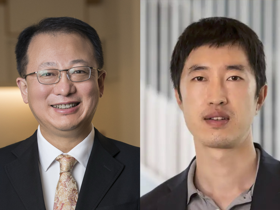
Co-principal investigator Timothy Holy, PhD, the Alan A. and Edith L. Wolff Professor of Neuroscience, and Ulugbek Kamilov, PhD, an assistant professor of electrical and systems engineering at Washington University’s McKelvey School of Engineering, aim to create an integrated software pipeline that will take in big data and turn out a manageable dataset ready for analysis. The software, which is being written in Julia, a programming language that Holy helped build, will create easily analyzable datasets by combining all available measurements and then using deep-learning techniques to identify and classify the most biologically relevant parts.
Read the full story on the advanced imaging tool


