A one-two punch for photoacoustic imaging
Song Hu combines hardware and machine learning for precision imaging technique

Scientists use photoacoustic microscopy to measure various biomarkers in the body, but some of these measurements can be inaccurate due to limitations of the light-focusing beam that produces out-of-focus images. Engineers in the McKelvey School of Engineering at Washington University in St. Louis have designed a new technique that combines hardware and software innovations as a one-two punch that overcomes this obstacle with excellent accuracy.
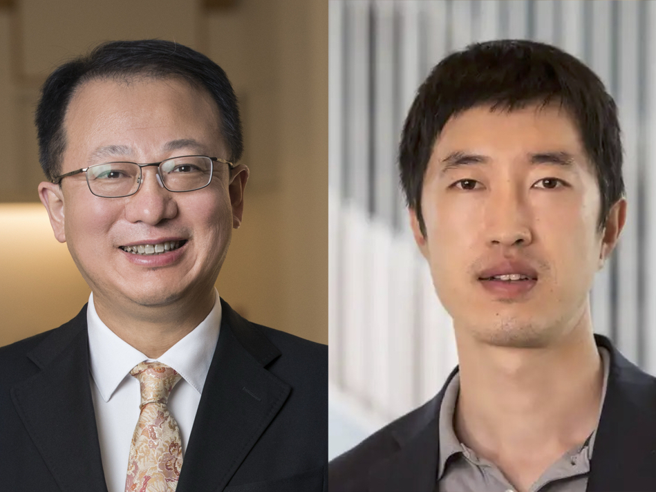
Song Hu, associate professor of biomedical engineering, along with Yifeng Zhou and Naidi Sun, members of his lab, combined a Bessel beam that propagates in the tissue in a diffraction-free manner with a conditional generative adversarial network-based deep learning model to obtain high-resolution photoacoustic images of hemoglobin concentration, blood oxygenation, and blood flow in the mouse brain with an extended depth of focus of about 600 microns. Compared with the conventional Gaussian-beam excitation based photoacoustic microscopy system, the combined method showed significantly higher quantitative accuracy.
Results of the research are published in IEEE Transactions on Medical Imaging, published online ahead of print July 5.
Hu and the team had several obstacles to overcome to reach their successful outcome, he said.
“The Bessel beam is able to focus into a nice spot over a much-extended depth range, but it has rings around it that compromise the image quality,” Hu said. “That’s where the machine learning approach comes in. We developed a machine learning algorithm to learn the difference between the Bessel beam-generated image and the image acquired in the focal plane of the Gaussian beam.”

While the Gaussian beam can provide a good, in-focus image over a small area of the brain cortex with a relatively flat surface, it cannot account for the uneven surface contour of the entire cortex. The Bessel beam combined with machine learning, however, provided a reliable measurement across the entire mouse cortex, the team found.
To train the conditional generative adversarial network, the team used data simultaneously acquired with the Gaussian beam and Bessel beam. Then, they compared the images acquired by the two beams to determine if the Bessel beam provided more accurate and in-depth information. Finally, they conducted imaging of the entire mouse cortex to show the benefits of Bessel beam photoacoustic microscopy over an uneven surface.
Hu said after sufficient learning, the machine learning algorithm is good enough to differentiate the true signal from the artifacts, or unwanted signals generated by side lobes of the Bessel beam and achieve the very large depth of focus offered by the Bessel beam. It is also free of artifacts.
Hu said this method could potentially be used in imaging of the brain and tumors, which both have uneven surfaces that are difficult to image with conventional light microscopy techniques.
Zhou Y, Sun N, Hu S. Deep Learning-powered Bessel-beam Multi-parametric Photoacoustic Microscopy. IEEE Transactions on Medical Imaging, early access online July 5, 2022. DOI: 10.1109/TMI.2022.3188739
This research was supported by funding provided by the National Institutes of Health (NS099261, NS 120481), the National Science Foundation (2023988) and the Chan Zuckerberg Initiative DAF.


