Mechanical strains, stresses behind traumatic brain injury to get closer look
Philip Bayly, collaborators to use imaging, modeling to look at waves in the brain

Naval warfighters may be exposed to explosions, impacts or high accelerations that increase their risk for traumatic brain injury. A team of researchers led by Philip Bayly at Washington University in St. Louis plans a comprehensive study of skull-brain mechanics using imaging, computer and preclinical models to study the strains and stresses of the brain induced by skull motion in these types of interactions.
Bayly, the Lee Hunter Distinguished Professor and chair of the Department of Mechanical Engineering & Materials Science in the McKelvey School of Engineering, and collaborators at the University of Delaware and Dartmouth College will conduct the research with a three-year, $700,000 grant from the Office of Naval Research. The work is expected to shed light on the ways that the magnitude, location and orientation of mechanical loading of the brain can influence the characteristics of a traumatic brain injury. Such injuries affect about 1.4 million people in the U.S. annually, both military personnel and civilians of all ages.
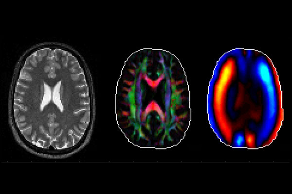
Bayly and his team will use several forms of imaging at the Neuroimaging Lab Research Center and the Small Animal Magnetic Resonance Facility in the Mallinckrodt Institute of Radiology at Washington University School of Medicine to study the behavior of the brain and skull in a preclinical model and to take measurements of the interactions between a helmet, the skull and the brain in healthy volunteers. Results of the imaging also will allow them to study the direction-dependent mechanical properties of brain tissue.
“We are looking at how shear waves move in the brain,” Bayly said. “A blast from an explosion creates a big pressure wave in air, which is transmitted into the skull potentially creating large, damaging, shear waves in the brain. In our experiments, we will generate small, non-injurious shear waves to probe how the brain might respond to blasts and other sources of skull motion.”
To understand shear waves, Bayly suggests visualizing gelatin (Jell-O) as a model for the brain. It is hard to change the volume of a block of gelatin but easy to deform it into a different shape. Shear waves are waves that deform the gelatin (or brain tissue), making them potentially more damaging, Bayly said.
Joining Bayly in the research at WashU is Ruth Okamoto, teaching professor in mechanical engineering & materials science, and Charlotte Guertler, a staff scientist. The computer modeling team will be led by Matthew McGarry, research assistant professor in the Thayer School of Engineering at Dartmouth College, and human imaging studies will be led by Curtis Johnson, co-principal investigator and assistant professor in the Department of Biomedical Engineering at the University of Delaware. Bayly, McGarry and Johnson are part of the ONR-funded Physics-based Neutralization of Threats to Human Tissues and Organs (PANTHER) Program, a nationwide consortium of researchers focused on the understanding, detection and prevention of traumatic brain injuries.
As part of the research, Bayly and his team will conduct a pilot study with a few healthy adult volunteers to describe how the standard military helmet would impact the motion transmission from a blast wave to the skull and to the brain. They will affix sensors to the subject’s front teeth through a custom mouthpiece, similar to those used by football players, to measure skull motion
“There are many ways that researchers are trying to mathematically estimate or calculate what is happening in the brain, but we want to provide hard data to inform computer models, then use that data to improve the design and development of protective devices,” Bayly said.


