Rupture of aorta to get closer look with specialized testing, imaging
Matthew Bersi will use a custom technique to study soft tissue rupture with a CAREER Award from the National Science Foundation

If you continuously inflate a balloon, eventually it will pop due to a material weakness somewhere in the balloon. Similarly, the aorta, the body’s largest artery, can develop weaknesses that can lead to a catastrophic rupture because of the mechanical demands of continuous blood flow over one’s lifetime. But researchers do not yet have the reliable tools to predict when and where a rupture may occur.
Matthew Bersi, assistant professor of mechanical engineering & materials science in the McKelvey School of Engineering at Washington University in St. Louis, will use pioneering optics-based mechanical testing and imaging techniques to study the aorta with a five-year, $575,000 CAREER Award from the National Science Foundation. CAREER awards support junior faculty who model the role of teacher-scholar through outstanding research, excellence in education and the integration of education and research within the context of the mission of their organization. One-third of current McKelvey Engineering faculty have received the award.
“The heart beats every second and is part of a system that is well designed for resistance against fatigue,” Bersi said. “In cases where there is damage to the aorta, changes in blood pressure with each beat of the heart can make the vessel vulnerable to failure, but this process is difficult to study and hard to predict since it is dependent upon the local mechanical environment of the tissue.”
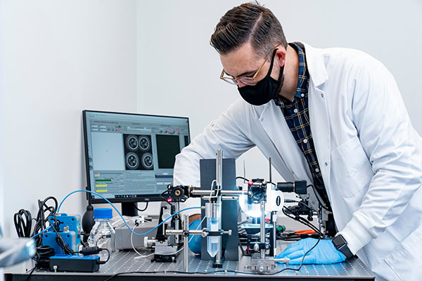
Bersi co-developed a unique optical system that uses a series cameras and mirrors to enable simultaneous visualization all the way around a tissue sample. Only two of these systems exist in the world. This system allows researchers to reconstruct the local mechanical environment around defects, or areas of weakness which may eventually rupture, in a model that can be more easily studied than in humans.
“We will place tissues that have vulnerability into our mechanical testing system, which allows us to measure the local mechanical properties,” Bersi said. “We will inflate these vessels, and they will eventually pop, or fail, and our system will allow us to see all around the vessel while inflating so we can see where and when it fails and measure the mechanical environment just before failure.”
In addition, Bersi and his team will use optical coherence tomography (OCT), a noninvasive imaging method that detects differences in how tissue refracts light and can acquire high-resolution 3D images with a depth of up to 1 to 2 millimeters in less than a minute.
“OCT is great for imaging soft tissues because light scatters well in collagen, the main structural protein in body, and allows us to see the tissue structure,” Bersi said. “It will allow us to look locally at the defects we put into the vessel wall and see how they respond to pressure and fatigue testing to determine how this damage propagates and contributes to failure.”
This method of research can be applied to the body’s other soft tissues in addition to the aorta, Bersi said. He plans to create an open-source computational model of aortic rupture based on his findings and will share the construction and operation of the technology he co-developed.
As part of the grant, Bersi will create outreach programs for students in kindergarten through sixth grade, as well as undergraduate and graduate students at WashU, that would include new coursework and hands-on demonstrations related to optics and mirrors, material structure and function and cardiovascular health. In addition, he plans to create a biological physics course for students interested in STEM in the university’s Prison Education Project.


