A true visionary
From amateur photography to professional imaging technology, Chao Zhou has developed the picture-perfect career
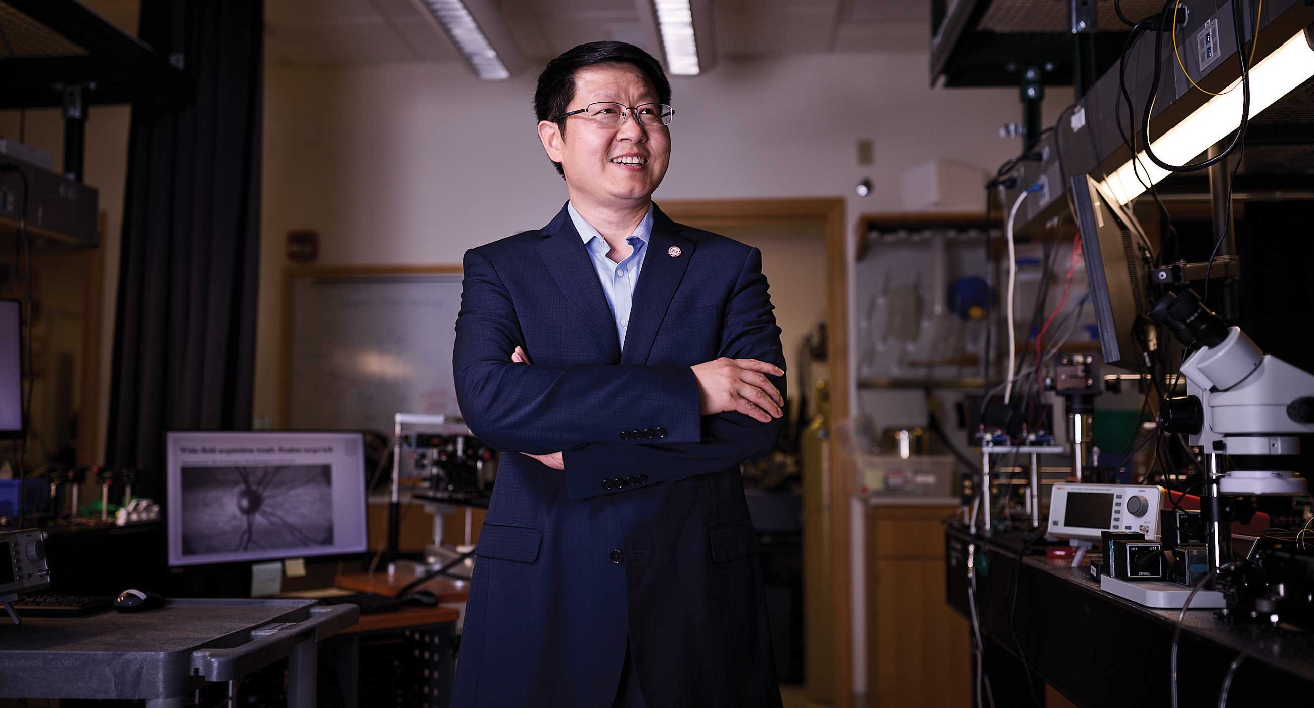
With a love of capturing images of the world around him, Chao Zhou joined a photography club when he was an undergraduate student studying physics at Peking University in China.
“I enjoyed taking pictures and sharing them with others,” says Zhou, now professor of biomedical engineering at the McKelvey School of Engineering. “When I moved to the University of Pennsylvania to work on my doctorate, it felt natural to focus on something related to images.”
Zhou’s PhD project involved the use of diffuse optical imaging to look at the effects a traumatic brain injury had on cerebral blood flow and oxygenation, providing critical information for clinical intervention.
“It was eye-opening,” he says. “My career in imaging technology started as a hobby, and then I realized it’s also useful for medicine and research. I’m able to contribute to the advancement of the technology and try to make it even better.”
With this technology, we don’t have to worry about people moving their head or blinking. Moreover, the current clinical OCT system is bulky and costly, so only major hospitals and clinics can afford it. We want to change that.
— Chao Zhou
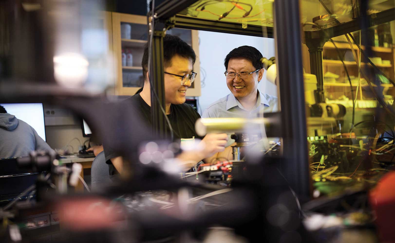
Jiawei Meng, research project manager, and Chao Zhou work on developing chips.
Seeing the light
After earning a doctorate, Zhou headed to the Massachusetts Institute of Technology for postdoctoral training and learned about a new type of imaging technology – optical coherence tomography (OCT), a noninvasive imaging technique that uses light to create cross-sectional high-resolution images of tissue. OCT is primarily used in ophthalmology to diagnose and monitor eye diseases and has been very successful at preventing vision loss in patients. Zhou continued working with OCT as he began his academic career at Lehigh University, where he was a founding member of the Department of Bioengineering.
“OCT is an exciting field, and we continue to improve the technology and add more functionality,” he says.
Early OCT devices, developed in the 1990s, were difficult to use and took several minutes to take just one picture. But in the early 2000s, a breakthrough in technology greatly improved their speed, and devices could then take one picture in as quickly as a fraction of a second.
“This allowed a wider adoption of the technology because high-quality images obtained from the patients led to a more accurate diagnosis,” Zhou says.
We want to use integrated photonics, so we can do everything on the same chip – guide the light to where we want and process optical signals that can be used to generate the image.
— Chao Zhou
Zhou and his lab are working on space-division multiplexing OCT, which could take multiple high-resolution images of the eye 10 times faster than traditional scanners.
Getting the full picture
While technology had increased the speed of OCT, it hadn’t yet fully met clinical needs. Initially, OCT could only take a few cross sections of the retina. When patients returned for follow-up imaging, it was difficult to align the new images with those from their first visit – making it hard to accurately track disease progression. This led to a new technology that could scan the entire eye to get volumetric data.
“With a high-density cube scan, you can look at the same position every time a patient comes back for imaging,” Zhou says. “It was much easier to align the images and track disease progression.”
One downside of the new technology? Getting a full scan of the eye took several minutes – a long time for people to sit still and not blink. Some young patients even needed to be sedated for the procedure to get high-quality images.
Zhou wanted to find a way to scan much faster to improve data quality, so he and his research team developed space-division multiplexing OCT. This new technique could take multiple high- resolution images 10 times faster than traditional scanners.
“With this technology, we don’t have to worry about people moving their head or blinking,” he says. “Moreover, the current clinical OCT system is bulky and costly, so only major hospitals and clinics can afford it. We want to change that.”
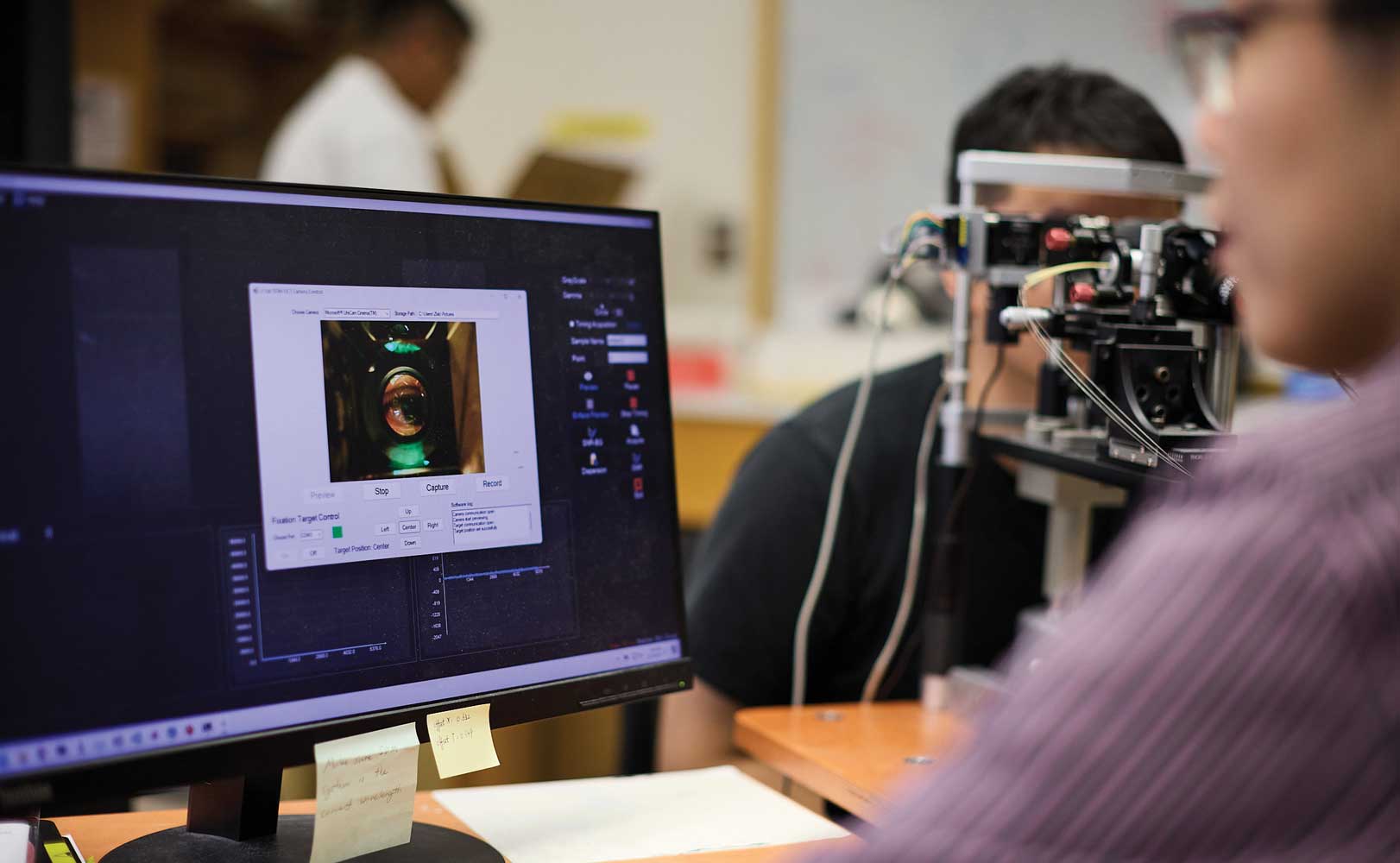
Zhou's lab uses this model of a human eye in its research.
Envisioning a solution
Zhou recently received an up to $20 million contract from the Advanced Research Projects Agency for Health (ARPA-H) — the first of its kind awarded to WashU — to develop a portable OCT device that is low cost, easy to use, and more than 30 times faster than existing systems. Zhou and his team will use photonic integrated circuits and custom-designed electronic integrated circuits to create their new device.
One example is the computer chip. Over the years, the size of a computer chip has shrunk to only a few centimeters and contains billions of transistors. However, these chips use silicon wafers, which are difficult for OCT imaging light to travel through.
“We want to use integrated photonics, so we can do everything on the same chip – guide the light to where we want and process optical signals that can be used to generate the image,” Zhou says.
By using the semiconductor industry’s advancements in complementary metal-oxide-semiconductor (CMOS) processes to create the chip, the team can streamline manufacturing and lower costs. They anticipate that their new technology could also be applied to cardiology, dermatology, dentistry, endoscopy, gynecology and urology.
Zhou credits WashU’s collaborative environment with helping him bring his technology from the lab into clinics. “WashU encourages people to work together,” he says. “I’ve established close collaborations with colleagues in McKelvey Engineering and with world-class physicians at the School of Medicine. When creating biomedical devices, you don’t want them to stay in the lab – you want them to go into clinics to benefit patients. WashU allows me to do this.”