Imaging technique can reduce benign breast biopsies by 25%
Ultrasound-guided diffuse optical tomography reduces breast biopsies in WashU study
In breast cancer, a biopsy is the only diagnostic procedure that can determine if a suspicious lump or area is cancerous. However, in the U.S., between 75% and 80% of breast biopsies are benign.
A team of researchers and physicians led by Quing Zhu, the Edwin H. Murty Professor of Engineering in the McKelvey School of Engineering at Washington University in St. Louis, and Debbie Bennett, MD, the Ronald and Hanna Evens Endowed Chair of Women’s Health at Barnes-Jewish Hospital and chief of breast imaging for WashU Medicine Mallinckrodt Institute of Radiology (MIR), have conducted a double-blind clinical trial involving 226 patients, utilizing ultrasound-guided diffuse optical tomography technology, to assess if the technology could reduce benign breast biopsy rates.
The study found that using this technology can reduce unnecessary breast biopsy rates by nearly 25% with a low false negative rate below 2% — well within the guideline of the American College of Radiology on standard of care biopsy decision risk.
Results of the research were published online in Breast Cancer Research Dec. 31, 2025. It was supported by a nearly $2 million grant from the National Cancer Institute of the National Institutes of Health.
Zhu and her team have pioneered this noninvasive imaging technique that uses near-infrared light to create 3D pictures of tissues, revealing functional differences between normal and cancerous tissue of blood vascular contrast and oxygen levels. The technology, when combined with standard of care ultrasound evaluation, can significantly triage low-risk breast lesions regarding biopsy. Considering that there are more than 1 million annual breast biopsies in the United States each year, this 25% biopsy reduction has a significant impact on saving health care cost and patient anxiety, Zhu said.
“With diffuse optical tomography, the resolution is lower because light is scattered in the tissue,” Zhu said. “Because of the ultrasound localization, we avoid the uncertainty and improve diffuse optical tomography.”
Zhu said this study is the first of its type in the diffuse optical imaging field with a large number of patients and several radiologists involved.
“The radiologists did not know our results, but evaluated each patient based on standard of care to get a risk evaluation and determine whether to do a biopsy,” she said. “Then they used our breast lesion hemoglobin maps and oxygenation saturation levels to decide upgrading or downgrading the lesion regarding a biopsy decision.”
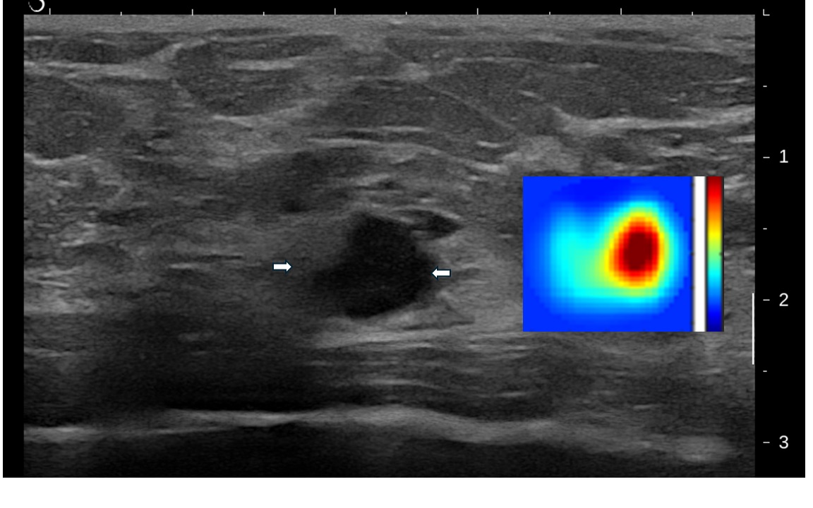
The team studied 226 patients with a breast lesion with a likelihood of malignancy and imaged them with ultrasound-guided diffuse optical tomography prior to their standard of care ultrasound-guided biopsy. The ultrasound-guided diffuse optical tomography provided total hemoglobin concentration maps and blood oxygen saturation levels to the study radiologists for reconsidering their biopsy decision. Hemoglobin, which is carried by red blood cells, absorbs the near-infrared light. A higher level of hemoglobin raises suspicion for cancer. Blood oxygen saturation, or the amount of oxygen in the blood, is another biomarker for indicating tumor metabolism. A lower level of blood oxygen saturation is indicative of suspicion for cancer.
The total hemoglobin concentration of patients with a malignant tumor was significantly higher than that of all benign lesions, the researchers found. The blood oxygen saturation was significantly lower in malignant lesions than in benign lesions. In addition, total hemoglobin concentration was significantly higher in grade 3 cancerous lesions than in grades 1 and 2, while the blood oxygen saturation was significantly lower. While the blood oxygen saturation of cancerous lesions smaller than 2 centimeters was not significantly different from that of cancerous lesions larger than 2 centimeters, the blood oxygen saturation differed significantly between the two groups.
In total, they found 70 invasive carcinomas, seven ductal carcinomas in situ, nine high-risk lesions and 140 benign lesions. Integrating total hemoglobin concentration and blood oxygen saturation derived from ultrasound-guided diffuse optical tomography with the classification assessment from the radiologists reduced the number of unnecessary benign biopsies by 24.54%.
Zhu said the team is looking to further reduce the system cost, optimizing AI-tools developed for data processing, imaging reconstruction, and dual-modality based lesion diagnosis, and commercialize the technology so it could be used at the bedside in a clinical setting.
The study team consisted of physicians of Steven P. Poplack, MD, professor of radiology (breast imaging) at Stanford University; Ian Hagemann, MD, PhD, professor in the Department of Pathology at WashU Medicine; Jaimee Mannix, MD, assistant professor of radiology, Kimberly Wiele, MD, professor of radiology, and Megan Luther, clinical research coordinator II, all from MIR; and Jingqin Luo, professor of surgery, Division of Public Health Sciences, at WashU Medicine.
Zhu Q, Bennett D, Hagemann IS, Mannix J, Wiele K, Luther M, Luo J, Poplack SP. Ultrasound-guided diffuse optical tomography: An adjunct to ultrasound that can reduce unnecessary breast biopsies. Breast Cancer Research, published online Dec. 31, 2025. DOI: https://doi.org/10.1186/s13058-025-02206-3
Funding for this research was provided by the National Cancer Institute of the National Institutes of Health (R01CA228047).
Click on the topics below for more stories in those areas
Back to NewsFaculty in this story

Quing Zhu
In the Media
The Conversation: About 80% of breast cancer biopsies turn out benign – new imaging tool promises clearer diagnoses and fewer biopsies