

A painless punch with a microneedle patch could detect kidney disease earlier
Srikanth Singamaneni’s lab creates metal-organic framework-protected microneedle patch resilient to heat, storage time
Kidney disease is silent in its early stages, progressing without symptoms until the disease is advanced. Researchers in the McKelvey School of Engineering at Washington University in St. Louis, in collaboration with researchers at Texas A&M University and Washington University School of Medicine, have been developing a minimally invasive method to improve diagnosis in earlier stages.
New research from the lab of Srikanth Singamaneni, the Lilyan & E. Lisle Hughes Professor in the Department of Mechanical Engineering & Materials Science, shows the early success of a microneedle patch that can be applied on the skin to quickly and safely capture biomarkers from the biofluid in the skin, or dermal interstitial fluid, and quantify them accurately on the patch. The patch detected early signs of kidney injury and may one day support home or point-of-care monitoring without requiring refrigeration.
Results of their research were published online Aug. 5 in Advanced Materials. It is the first study to show that encapsulating biomolecules on microneedles preserved their biological functions.
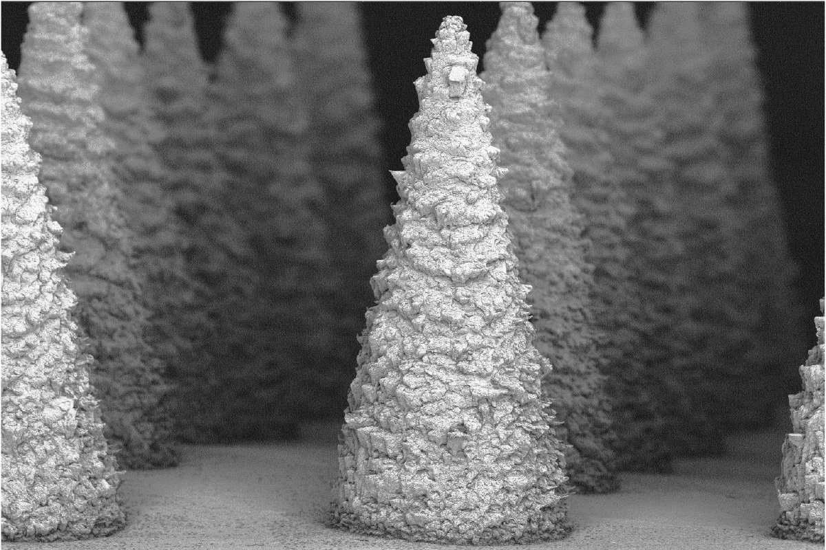
The research team, which includes Yixuan Wang, a doctoral student in Singamaneni’s lab, created microneedles coated with a metal-organic framework (MOF) that can sample interstitial fluid in the skin. The microneedles are coated with zeolitic imidazolate framework-8, which creates a shell that detects and preserves the neutrophil gelatinase-associated lipocalin (NGAL) antibodies, an early biomarker of acute kidney injury. The MOF shell preserved the antibodies for up to four weeks at 50 C (122 F) without refrigeration.
“This metal-organic framework encapsulation is a simple and highly effective way to create microneedle sensors that are resilient to environmental challenges and provide a scalable path to minimally invasive biosensing for at-home or remote health monitoring without needing cold chain,” Singamaneni said.
NGAL is a glycoprotein that increases in the blood within hours of a kidney injury and has become a clinically validated biomarker for kidney damage. Because it requires a lab setting, drawing blood with a needle and cold chain logistics, it has not been useful in home-based or resource-limited settings.
Previously, Singamaneni and collaborators established polystyrene-based microneedle patches combined with plasmonic fluor as nanolabels for ultrasensitive detection of low-abundance protein biomarkers. Adapting that technology required them to create a biosensor with high sensitivity and a broader range, as well as addressing cold-chain logistics.
In the new research, the microneedle-based sensors, shaped like traffic cones, penetrate only the outer layers of skin, which avoids pain, bleeding and the need for trained personnel to use them. Adding metal-organic frameworks to the microneedles stabilizes the biomolecules in high-heat settings.
In collaboration with Limei Tian’s lab at Texas A&M University and the lab of Maggie Chen, MD, PhD at WashU Medicine, the team tested the microneedle patch in a mouse model as well as in humans. Researchers applied the patch to the mouse skin for 1 minute to capture and measure NGAL. The NGAL levels measured via the microneedle patch qualitatively matched those in the blood samples.
In tests on humans, the team applied the patch for 5 minutes. After it was removed, there was no bleeding, and the skin fully recovered within 30 minutes. While the NGAL concentration captured from the microneedle patch was slightly lower than that from blood plasma, the two measurements were closely correlated.
The next steps, Singamaneni said, are to adapt the microneedle method to other proteins, metabolites and nucleic acids to broaden its scope of decentralized diagnostics. In addition, more research is needed to characterize protein markers in interstitial fluid relative to blood and to shed light on the dynamics of their concentration and diffusion kinetics to inform clinically actionable thresholds.
Wang Y, Liu Y, Debnath A, Li C, Want Z, Kondepati G, Balmoori S, Morrissey JJ, Chen YM, Tian L, Singamaneni S. Metal-Organic Framework-Preserved Thermostable Microneedle Patch for Minimally Invasive Detection and Monitoring of Kidney Dysfunction. Advanced Materials, published online Aug. 5, 2026. DOI: https://doi.org/10.1002/adma.74423
This research was supported by funding from the National Science Foundation (CBET 2224610 and CBET 560 2316285), the National Institutes of Health (R21DK131557A1, R35 GM147568, R01 2DK105056-6 and R56DK138158A1), the Congressionally Directed Medical Research Programs (HT94252310996 and W81XWH-19-1-0320), and VA Merit (I01BX006401A1).
Singamaneni and Jeremiah J. Morrissey are the inventors of the plasmonic-fluor technology, which has been licensed by the Office of Technology Management at Washington University in St. Louis.
Click on the topics below for more stories in those areas
Back to NewsFaculty in this story