
Quick optical biopsy could be early detection method for endometrial cancer
Quing Zhu, WashU Medicine collaborators combine optical coherence tomography and machine learning for rapid, accurate test
Endometrial cancer is the most common gynecologic cancer, with more than 69,000 cases diagnosed in the U.S. in 2025 and increasing up to 3% annually. Diagnosis requires an often painful and invasive biopsy that carries a risk of false negatives. A multidisciplinary research team at Washington University in St. Louis and Siteman Cancer Center, based at Barnes-Jewish Hospital and WashU Medicine, is looking to a fast, safe and noninvasive imaging method combined with machine learning for an accurate detection and diagnosis of precancerous lesions and early cancers.
The team, led by Quing Zhu, the Edwin H. Murty Professor of Engineering in the McKelvey School of Engineering at Washington University in St. Louis, conducted an initial investigation using optical coherence tomography (OCT), which detects differences in how tissue reflects light and acquires high-resolution 3D images with a depth of up to 1 to 2 millimeters. With a custom catheter probe developed in Zhu’s lab, the team took images of the entire endometrial cavity in less than 3 minutes, creating an optical biopsy. It is the first catheter-based, 3D OCT imaging study that integrated optical functional, structural and radiomic features for endometrial assessment. Results of the research were published in npj Imaging June 3, 2026.
To obtain images from patient tissues, the team collaborated with WashU Medicine physicians led by Lindsay Kuroki, MD, associate professor of obstetrics & gynecology, and Ian Hagemann, MD, PhD, professor of pathology & immunology and of obstetrics & gynecology. They, along with Zhu, are research members at Siteman Cancer Center, where Kuroki also treats patients. The team acquired OCT images from 57 post-hysterectomy uteri in 2025. Of these, 34 contained high-risk precancerous lesions or early-stage cancers.
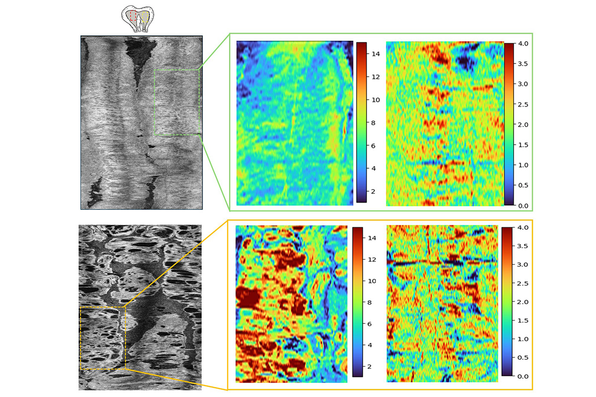
The 3D OCT images provided a close view of tissue microstructure and optical properties, revealing clear differences among normal endometrium, benign endometrium, high-risk precancerous lesions, and endometrial cancer at different stages.
First authors Sanskar Thakur, a doctoral student in Zhu’s lab, and Yixiao Lin, who earned a doctorate in biomedical engineering from WashU in 2025, developed an imaging feature extraction pipeline and a machine learning model to categorize the results into two groups of normal and benign, and pre-cancer and cancer using 26 extracted imaging features. Their model achieved an exploratory sensitivity of 94% and specificity of 87%.
“Current endometrial biopsy practice has an estimated false-negative rate of about 10% (approximately 90% sensitivity), largely due to sampling limitations and interpretive variability,” Zhu said. “With our three-dimensional OCT imaging system combined with machine learning, we can image the entire endometrial cavity in 2 to 3 seconds and may have a potential to achieve higher sensitivity than random biopsy sampling.”
“There is currently no reliable screening for endometrial cancer,” said coauthor David Mutch, the Ira C. and Judith Gall Professor and vice chair of obstetrics & gynecology at WashU Medicine, a Siteman research member and principal investigator of the National Cancer Institute-funded Route 66 Endometrial Cancer Specialized Program of Research Excellence (SPORE) grant. “This technology, developed by Dr. Zhu and her colleagues, should allow us to better screen for this cancer and at a minimum catch it much earlier in its development,” Mutch added. “This is really novel, cutting-edge technology.”
Going forward, Zhu said the team plans to evaluate the catheter in live patients to demonstrate the translational potential of the AI-assisted OCT technology.
Thakur S, Lin Y, Xu J, Nie H, Badwan S, Wang L, Sanders BE, Thaker PH, Hagemann AR, McCourt CK, Khabele DM, Powell MA, Mutch DG, Kuroki LM, Hagemann IS, Zhu Q. Optical coherence tomography enables optical biopsy of endometrial tissue for early cancer detection. npj Imaging, June 3, 2026, https://doi.org/10.1038/s44303-026-00160-z.
This work was funded by the Developmental Research Program (DRP) of the NCI Route 66 Endometrial Cancer SPORE (5P50CA265793-03). Partial support for this work was provided by the NCI (R01CA237664) and the NIBIB (R01EB034398).