
Tiny feet on cells sense defects, stall migration to heal wounds
Finding in Amit Pathak’s lab impacts injury sensing by cells
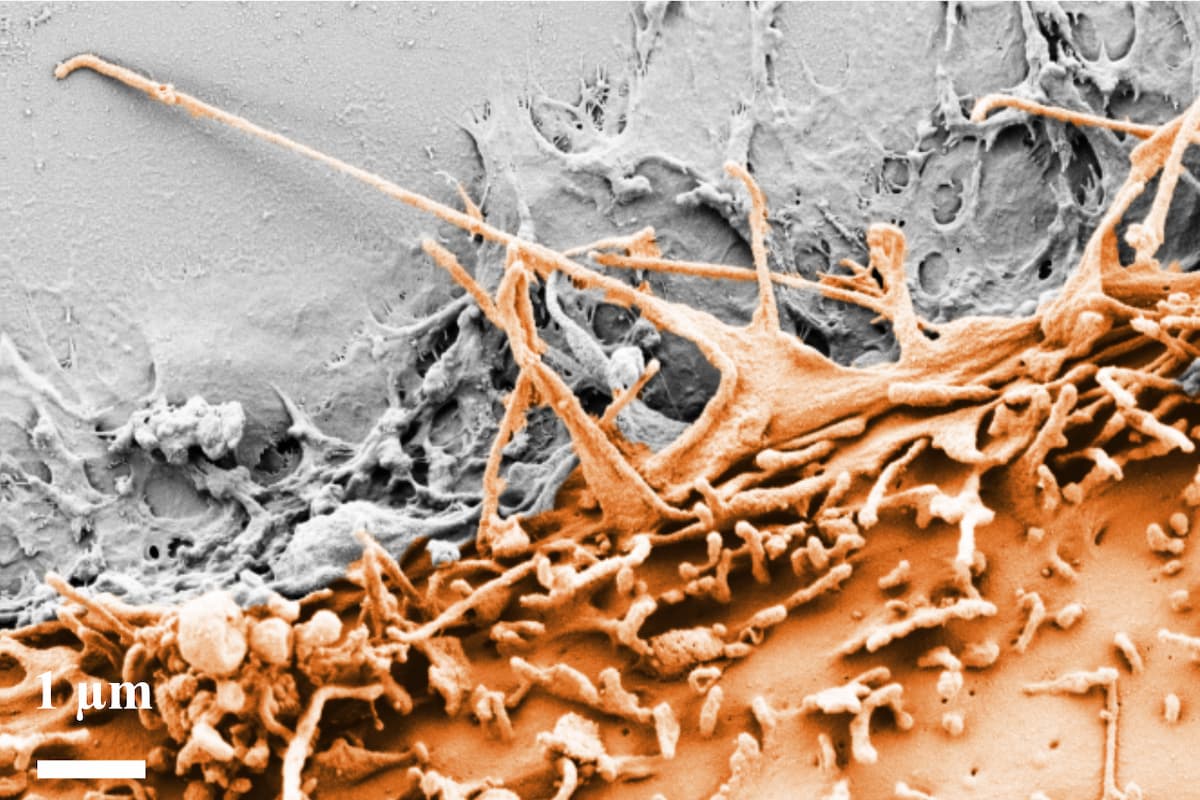
Cells travel through the body both individually and in collective groups during development, in wound healing and in diseases, such as cancer. New research from the McKelvey School of Engineering at Washington University in St. Louis shows that cells can sense even the smallest defects, or micro-injuries, in the surface below that which they are traveling and stall long enough to begin the healing process.
Amit Pathak, professor of mechanical engineering & materials science, and members of his lab, including Hannah Zmuda, who earned a doctorate in biomedical engineering from McKelvey Engineering in 2025, found that the tiny feet on the leading edge of a group of cells can sense a defect of only a few microns in a membrane below the surface of the extracellular matrix, which supports cell growth and provides structure when tissues regenerate. Their results were published July 31, 2026, in Cell Reports.
Groups of cells travel along the extracellular matrix (ECM) that has a basement membrane, a protective layer made from collagen IV that acts as a tissue barrier for organs, and the interstitial matrix, which is made of collagen I and sits below the basement membrane. These cells run along the ECM with tiny feet called filopodia, which are so sensitive that they can determine the smallest of defects in the basement membrane and stop for up to eight hours — but only when collagen IV was present.
“The filopodia were something we specifically were not looking for, but they turned out to be the things that control this sensing of these tiny defects in extracellular surfaces,” Pathak said.
Interestingly, when the filopodia sense collagen I or stiffer ECMs, they kept moving over the defect, Pathak said, showing that a difference in stiffness is important.
“In cancer, the basement membrane gets degraded completely, so the cells have access to collagen type 1, which is the more native tissue,” Pathak said. “In tumor invasion, you want them to notice the gap and stop, because stalling is good in cancer. If the cells don't notice small wounds and keep going, that means there was no healing done. If they stall and deposit new extracellular matrix, they heal the wound and then move on.”
Zmuda, who had a prestigious National Science Foundation Graduate Research Fellowship, and the team also found that the environment around the cells affects how strongly the stalling spreads, particularly how stiff the surrounding material is and how osmolar the fluid medium is, in addition to the type of collagen that is present.
“When we culture cells in the body, the fluid is not all the same,” Pathak said. “There are differences in salt, sugar and water, which has a huge effect on osmolality. This changes cell migration and defect sensing. The membrane changes their feet, and the filopodia change.”
The team conducted their research with human mammary epithelial cells, Madin-Darby Canine Kidney cells and primary zebrafish keratinocytes from fish scales. They created the defects in the hydrogels with laser ablation, then imaged the movement using atomic force microscopy.
Pathak said the findings show the importance of studying how multiple extracellular cues, including protein types, stiffness and osmotic conditions, alter subcellular mechanics and multicellular collective migration. Better understanding these independent influences will increase knowledge of basic biological processes in wound healing, development and tumor invasion, he said.
Zmuda H, Barra Avila D, Lee P-H, Walter C, Pathak A. Microscale matrix defects suppress tension-dependent protrusions and stall collective cell migration. Cell Reports. July 31, 2026, DOI: 10.1016/j.celrep.2026.117754
This research was supported by funding from the National Institutes of Health (R35GM156571) and the National Science Foundation Graduate Research Fellowship Program (DGE-2139839 and DGE-1745038).