WashU researchers develop first human model for form of inherited heart disease
Engineered tissues will help improve understanding of DRM, other cardiomyopathies
Inherited cardiomyopathies — diseases of the heart muscle, which cause the heart to have a harder time pumping blood to the rest of the body — are the leading cause of heart failure in young adults. Desmin-related cardiomyopathy (DRM) is one such familial disease that affects approximately 1 in 2,000 individuals worldwide. No disease-specific therapies exist for DRM, with patients receiving only generic heart failure medicines.
DRM is caused by mutations in the genes for desmin — a muscle-specific intermediate filament protein — or its protein-folding chaperone, αB-Crystallin (CRYAB). These mutations are thought to cause desmin to misfold and form toxic clumps inside heart muscle cells.
Deadly variations of this disease have been difficult to study in part because existing mouse models of the disorder require massive overexpression of mutant CRYAB. Since part of how the toxic protein clumps form is through proteins interacting with one another, there are concerns about how useful a model that overexpresses the proteins is — after all, more protein would mean more interaction with other molecules.
Now the first human model of CRYAB-linked DRM has been developed by researchers at Washington University in St. Louis, using micro-engineered tissues and induced pluripotent stem cells that express mutant CRYAB at a concentration that closely mirrors actual levels in the human body.
“Up until now, progress has been hindered by reliance on experimental models with uncertain translational value,” said Nathaniel Huebsch, an associate professor of biomedical engineering at WashU McKelvey Engineering. “A human experimental system with physiologically relevant mutant protein expression has been a critical missing tool for understanding DRM pathogenesis and developing targeted therapies.”
In a study published Nov. 11 in Science Advances, Huebsch and an all-WashU research team describe the engineered tissue they developed and report on their findings about the role of impaired protein quality-control machinery in cardiomyopathy, as well as implications for potential treatments.
“This is a disease where desmin is aggregated and mislocalized. But the cardiomyocytes that you make in the lab from the human stem cells really don’t express this,” Huebsch said. Working collaboratively with researchers in the laboratory of Abhinav Diwan, MD, the Charlie W. Shaeffer, M.D. Professor in the John T. Milliken Department of Medicine at WashU Medicine, Huebsch’s graduate student Yasaman Kargar Gaz Kooh, first author of the study, found a way around this limitation. Other key McKelvey Engineering contributors included Guy Genin, Anastasia Khokhlova and Jonathan Silva.
Kargar used human induced pluripotent stem cells (iPSC) that had their genome edited to express the mutant version of CRYAB, and then used cardiomyocytes from these iPSC to create miniaturized engineered heart tissues. One of the features of the engineered heart tissues is that cardiomyocytes within these tissues experience mechanical tugging forces and have to tug against a resistive load — just like in a real heart.
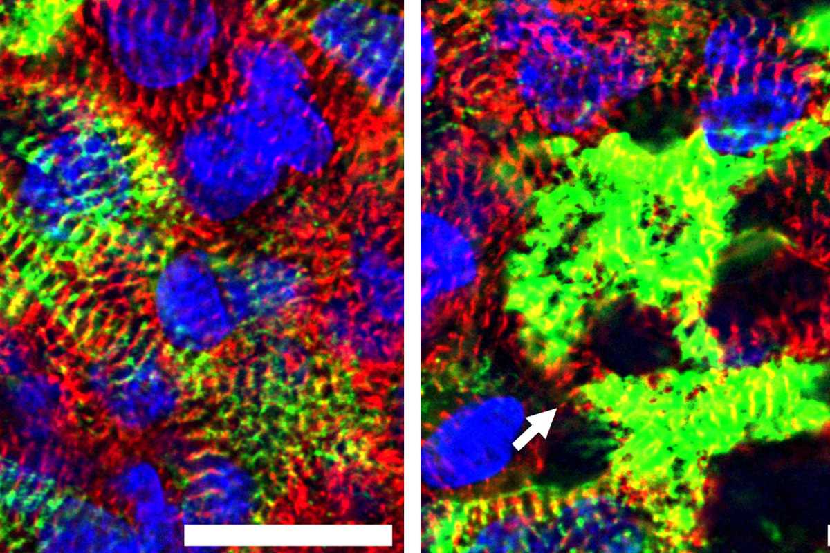
The mechanical forces within the engineered heart tissues turned out to be essential. Unlike heart muscle cells grown on flat surfaces in a lab dish, cells in these miniature tissues produced desmin and organized it much like they would in a real human heart. This allowed the researchers to recreate important features of the disease that are difficult to observe in traditional laboratory models.
“In our engineered tissue, perhaps because of the mechanical forces on the cells in the tissue, the cardiomyocytes do express that desmin, and we could start to learn about how the CRYAB mutation caused it to aggregate protein,” Huebsch said.
“We were able to look at healthy cells to see what the protein expression was, and where it was distributed. It goes to the Z-disc of the cardiomyocyte,” Kargar said. “Then, in the cells that have the mutation that causes the disease, we saw that it was a bit more aggregated. It was in clumps where, presumably, not only is it not having its intended function, but it may even be pulling other proteins with it.”